
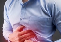
Um adulto do sexo masculino apontando para um contorno de fígado e pintou uma vesícula biliar em seu abdômen.
Definição e fatos do cálculo biliar
- Os cálculos biliares são "pedras" que se formam na vesícula biliar ou nos ductos biliares. Os tipos comuns de cálculos biliares são colesterol, pigmento preto e pigmento marrom.
- Os sintomas mais comuns de cálculos biliares são cólica biliar e colecistite; no entanto, geralmente, os cálculos biliares não causam sintomas.
- A dor da cólica biliar é um tipo muito específico que surge repentina ou rapidamente e atinge um pico em alguns minutos; no entanto, a dor pode variar em gravidade. O movimento não piora a dor.
- Outros sinais e sintomas de cólica biliar incluem:
- Náusea
- Dor comumente sentida na parte superior do abdômen
- Raramente, a dor pode ser sentida sob o esterno e é confundida com um ataque cardíaco ou angina (dor no peito).
- A cólica biliar geralmente tem um padrão que varia de pessoa para pessoa.
- Os cálculos biliares não causam intolerância a alimentos gordurosos, arrotos, distensão abdominal ou gases.
- As complicações dos cálculos biliares incluem colangite, gangrena da vesícula biliar, icterícia, pancreatite, sepse, fístula e íleo.
- O lodo da vesícula biliar está associado a sintomas e complicações de cálculos biliares; no entanto, como os cálculos biliares, o lodo geralmente não causa problemas.
- O melhor teste único para diagnosticar cálculos biliares é a ultrassonografia transabdominal. Outros exames incluem ultrassonografia endoscópica, colangiopancreatografia por ressonância magnética (CPRM), colecintilografia (exame HIDA), colangiopancreatografia retrógrada endoscópica (CPRE), exames de sangue hepático e pancreático, drenagem duodenal, colecistograma oral (OCG) e colangiograma intravenoso (IVC).
l>
- Os cálculos biliares são tratados principalmente com observação (sem tratamento) ou remoção da vesícula biliar (colecistectomia). Os tratamentos menos usados incluem esfincterotomia e extração de cálculos biliares, dissolução com medicamentos orais e litotripsia extracorpórea por ondas de choque (LECO). A prevenção de cálculos biliares de colesterol também é possível com medicamentos orais.
- Sintomas de cálculos biliares devem interromper a seguinte colecistectomia. Caso contrário, é provável que os cálculos biliares tenham sido deixados nos dutos, há um segundo problema nos dutos biliares ou os sintomas são causados por outro problema.
- Muitas recomendações alimentares foram feitos para a prevenção ou tratamento de cálculos biliares e para prevenir seus sintomas, mas nenhum deles se mostrou eficaz.
- Muitos remédios caseiros foram sugeridos para eliminar cálculos biliares, mas nenhum demonstrou ser eficaz
- As pesquisas contínuas visam descobrir os genes responsáveis pela formação dos cálculos biliares.
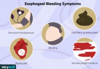
Sintomas de ataque da vesícula biliar
Os sintomas de um ataque da vesícula biliar incluem:
- dor no lado superior direito ou no meio do abdômen;
- a dor pode ser incômoda, aguda ou em cólica;
- a dor geralmente começa de repente;
- a dor é constante e pode se espalhar para as costas ou para a área abaixo da omoplata direita.
Leia mais sobre os sintomas de ataque da vesícula biliar »

Ilustração do sistema digestivo com cálculos biliares e close-up de cálculos biliares na vesícula biliar uma pedra que também passou para o ducto cístico.
O que são cálculos biliares? Como eles se formam?
Os cálculos biliares (muitas vezes mal escritos como cálculos biliares) são pedras que se formam na bílis (bile) dentro da vesícula biliar. (A vesícula biliar é um órgão em forma de pêra logo abaixo do fígado que armazena a bile secretada pelo fígado.) Os cálculos biliares atingem um tamanho entre um décimo de sexto de polegada e vários centímetros.
- Bile é um líquido aquoso produzido pelas células do fígado que é importante para digerir alimentos no intestino, principalmente gordura, e eliminar substâncias tóxicas do corpo.
- As células do fígado secretam a bile em pequenos canais dentro do fígado, chamados de canalículos.
- A bile flui através dos canalículos e em ductos coletores maiores dentro do fígado, chamados de ductos biliares intra-hepáticos.
- A bile então flui através dos ductos biliares intra-hepáticos fundidos para fora do fígado como ductos biliares extra-hepáticos (fora do fígado) (primeiro para os dois ductos biliares hepáticos, depois para o ducto hepático comum único e, finalmente, após o ducto hepático comum ducto é unido pelo ducto cístico proveniente da vesícula biliar, no ducto biliar comum.
Do ducto biliar, a bile pode fluir de duas direções diferentes.
- A primeira direção é através do ducto biliar comum e diretamente no intestino, onde a bile se mistura com os alimentos e promove a digestão dos alimentos. Ao mesmo tempo, substâncias tóxicas que são removidas do sangue pelo fígado são eliminadas no intestino.
- A segunda direção é para uma ramificação do ducto biliar comum, o ducto cístico, e daí para a vesícula biliar.
Uma vez na vesícula biliar, a bile é concentrada pela remoção (absorção) de água. Durante uma refeição, o músculo que compõe a parede da vesícula biliar se contrai e comprime a bile concentrada na vesícula biliar de volta através do ducto cístico para o ducto biliar comum e depois para o intestino. (A bile concentrada é muito mais eficaz para a digestão do que a bile não concentrada que vai do fígado direto para o intestino.) O momento da contração da vesícula biliar - durante uma refeição - permite que a bile concentrada da vesícula biliar se misture com os alimentos.
Os cálculos biliares geralmente se formam na vesícula biliar; no entanto, eles também podem se formar em qualquer lugar onde haja bile - nos ductos intra-hepáticos, hepáticos, biliares comuns e císticos.
Os cálculos biliares também podem se mover na bile, por exemplo, da vesícula biliar para o ducto cístico ou comum.

Um homem sente dor de cólica biliar. O sintoma mais comum de cálculos biliares é a cólica biliar.
Quais são os sinais e sintomas de cálculos biliares? Eles causam dor?
A maioria das pessoas com cálculos biliares não apresenta sinais ou sintomas e não tem conhecimento de seus cálculos biliares. (Os cálculos biliares são "silenciosos".) Esses cálculos biliares geralmente são encontrados como resultado de testes (por exemplo, ultra-som ou raios-X do abdômen) realizados durante a avaliação de outras condições médicas que não os cálculos biliares. Os sintomas podem aparecer mais tarde na vida, no entanto, após muitos anos sem sintomas. Assim, em um período de cinco anos, aproximadamente 10% das pessoas com cálculos biliares silenciosos desenvolverão sintomas. Uma vez que os sintomas se desenvolvem, é provável que continuem e, muitas vezes, piorem.
Quando ocorrem sinais e sintomas de cálculos biliares, eles quase sempre ocorrem porque os cálculos biliares obstruem os ductos biliares.
O sintoma mais comum de cálculos biliares é a cólica biliar. A cólica biliar é um tipo muito específico de dor, ocorrendo como sintoma primário ou único em 80% das pessoas com cálculos biliares que desenvolvem sintomas. A cólica biliar ocorre quando os ductos biliares (ductos císticos, hepáticos ou ductos biliares comuns) são subitamente bloqueados por um cálculo biliar. A obstrução de progressão lenta, a partir de um tumor, não causa cólica biliar. Atrás da obstrução, o líquido se acumula e distende os ductos e a vesícula biliar. No caso de obstrução do ducto hepático ou do ducto biliar comum, isso se deve à secreção contínua de bile pelo fígado. No caso de obstrução do ducto cístico, a parede da vesícula biliar secreta líquido na vesícula biliar. A distensão dos ductos ou da vesícula biliar causa cólica biliar.
Caracteristicamente, a cólica biliar surge repentinamente ou aumenta rapidamente para um pico em alguns minutos.
- É uma dor constante; ela não vem e vai, embora possa variar em intensidade enquanto está presente. A TI não é como uma cãibra.
- Dura de 15 minutos a 4-5 horas. Se a dor durar mais de 4-5 horas, significa que uma complicação - geralmente colecistite - se desenvolveu.
- A dor geralmente é intensa, mas o movimento não piora a dor. Na verdade, os pacientes com cólica biliar costumam andar ou se contorcer (torcer o corpo em diferentes posições) na cama tentando encontrar uma posição confortável.
- A cólica biliar geralmente é acompanhada de náusea.
- Mais comumente, a cólica biliar é sentida no meio da parte superior do abdômen, logo abaixo do esterno.
- O segundo local mais comum de dor é o abdome superior direito, logo abaixo da margem das costelas.
- Ocasionalmente, a dor também pode ser sentida nas costas, na ponta inferior da escápula, no lado direito.
- Em raras ocasiões, a dor pode ser sentida abaixo do esterno e é confundida com angina ou ataque cardíaco.
- Um episódio de cólica biliar desaparece gradualmente quando o cálculo biliar se desloca dentro do ducto, de modo que não está mais causando obstrução.
A cólica biliar é um sintoma recorrente. Uma vez que o primeiro episódio ocorre, é provável que haja outros episódios. Além disso, existe um padrão de recorrência para cada indivíduo, ou seja, em alguns indivíduos os episódios tendem a permanecer frequentes enquanto em outros são pouco frequentes. A maioria das pessoas que desenvolve cólica biliar não desenvolve colecistite ou outras complicações. Há um equívoco de que a contração da vesícula biliar é o que causa a obstrução dos ductos e a cólica biliar. Comer, mesmo alimentos gordurosos, não causa cólica biliar; a maioria dos episódios de cólica biliar ocorre durante a noite, muito depois do esvaziamento da vesícula biliar.
Os cálculos biliares são culpados por muitos sintomas que não causam. Entre os
sintomas que os cálculos biliares não causam são:
- dispepsia (incluindo inchaço abdominal e desconforto após comer),
- intolerância a alimentos gordurosos,
- arrotando e
- flatulência (passar gases ou peidar).

Ilustração mostrando cálculos biliares na vesícula biliar, bem como no ducto biliar comum distal. O ducto biliar comum tem uma parede muscular.
Você ainda pode ter sintomas de cálculos biliares depois que eles foram removidos?
A remoção da vesícula biliar (colecistectomia) deve eliminar todos os sintomas relacionados ao cálculo biliar, exceto em três situações:
- foram deixados cálculos biliares nos dutos,
- houve problemas com os ductos biliares, além de cálculos biliares, e
- os cálculos biliares não foram a causa dos sintomas.
A possibilidade de cálculos biliares nos ductos pode ser investigada com CPRM, ultrassom endoscópico e CPRE. Raramente, sintomas semelhantes a cálculos biliares podem ser causados por uma condição chamada disfunção do esfíncter de Oddi, discutida abaixo.
O ducto biliar comum tem uma parede muscular. Os últimos vários centímetros do músculo do ducto biliar comum imediatamente antes do ducto se juntar ao duodeno compreendem o esfíncter de Oddi. O esfíncter de Oddi controla o fluxo da bile. Como o ducto pancreático geralmente se une ao ducto biliar comum pouco antes de entrar no duodeno, o esfíncter também controla o fluxo de fluido do ducto pancreático. Quando o músculo do esfíncter se contrai, ele interrompe o fluxo de bile e fluido pancreático. Quando relaxa, a bile e o líquido pancreático fluem novamente para o duodeno, por exemplo, após uma refeição. O esfíncter pode ficar cicatrizado e o ducto é estreitado pela cicatriz. (A causa da cicatriz é desconhecida.) O esfíncter também pode entrar em espasmo intermitentemente. Em ambos os casos, o fluxo de bile e fluido pancreático pode parar de forma intermitente e abrupta, imitando os efeitos de um cálculo biliar causando cólica biliar e pancreatite.
O diagnóstico de disfunção do esfíncter de Oddi pode ser difícil de ser feito. O melhor teste diagnóstico requer um procedimento endoscópico com o mesmo tipo de endoscópio da CPRE. Em vez de preencher os dutos com corante, no entanto, a pressão dentro do esfíncter é medida. Se a pressão for anormalmente alta, são prováveis cicatrizes ou espasmo do esfíncter. O tratamento para a disfunção do esfíncter de Oddi é a esfincterotomia (descrita anteriormente). A medição das enzimas hepáticas e pancreáticas no sangue também pode ser útil no diagnóstico de disfunção esfincteriana.

Coleção de cálculos biliares de vários tamanhos e formas.
Como são os cálculos biliares?
Os cálculos biliares podem numerar de um a centenas, variando em tamanho de um milímetro a quatro ou cinco centímetros. Quando há um único ou apenas alguns cálculos biliares, eles tendem a ser redondos. Quando um número maior de cálculos biliares está presente, eles tendem a ser facetados devido à fricção de um cálculo biliar contra outro. Os cálculos biliares de pigmento marrom podem ser quebradiços e irregulares.
Os cálculos biliares passam?
Os cálculos biliares podem sair da vesícula biliar ou dos dutos, principalmente se forem pequenos. É a passagem de cálculos biliares que leva a muitas de suas complicações.
O que causa cálculos biliares? Quem os recebe?
Os cálculos biliares são comuns; ocorrem em aproximadamente 20% das mulheres nos EUA, Canadá e Europa, mas há uma grande variação na prevalência entre os diferentes grupos étnicos. Por exemplo, cálculos biliares ocorrem 1 ½ a 2 vezes mais comumente em escandinavos e mexicanos-americanos. Entre os índios americanos, a prevalência de cálculos biliares é superior a 80%. Essas diferenças provavelmente são explicadas por fatores genéticos (hereditários). Parentes de primeiro grau (pais, irmãos e filhos) de indivíduos com cálculos biliares são 1 ½ vezes mais propensos a ter cálculos biliares do que se não tiverem um parente de primeiro grau com cálculos biliares. Mais suporte para uma predisposição genética vem de estudos com gêmeos. Assim, entre pares de gêmeos não idênticos (que compartilham 50% de seus genes um com o outro), ambos os indivíduos em um par têm cálculos biliares 8% das vezes. Entre pares idênticos de gêmeos (que compartilham 100% de seus genes um com o outro), ambos os indivíduos têm cálculos biliares 23% das vezes.
Várias condições estão associadas à formação de cálculos biliares, e a maneira como eles causam cálculos biliares pode variar. (Veja os riscos de cálculos biliares.)
Os cálculos biliares de colesterol são compostos principalmente de colesterol. Existem dois outros processos que promovem a formação de cálculos biliares de colesterol
Cálculos biliares de colesterol
Existem vários tipos de cálculos biliares, e cada tipo tem uma causa diferente.
Cálculos biliares de colesterol
Os cálculos biliares de colesterol são compostos principalmente de colesterol. Eles são o tipo mais comum de cálculo biliar, compreendendo 80% dos cálculos biliares em indivíduos na Europa e nas Américas. O colesterol é uma das substâncias (produtos químicos) que as células do fígado secretam na bile. A secreção de colesterol na bile é um importante mecanismo pelo qual o fígado elimina o excesso de colesterol do corpo.
Para que a bile transporte colesterol, o colesterol deve ser dissolvido na bile. O colesterol é gordura, entretanto, e a bile é uma solução aquosa ou aquosa; as gorduras não se dissolvem em soluções aquosas. Para dissolver o colesterol na bile, o fígado também secreta dois detergentes, ácidos biliares e lecitina, na bile. Esses detergentes, assim como os detergentes para lavar louça, dissolvem o colesterol gorduroso para que ele possa ser transportado pela bile através dos dutos. Se o fígado secreta muito colesterol para o número de ácidos biliares e lecitina que secreta, parte do colesterol não permanece dissolvido. Da mesma forma, se o fígado não secretar ácidos biliares e lecitina suficientes, parte do colesterol não permanece dissolvido. Em ambos os casos, o colesterol não dissolvido gruda e forma partículas de colesterol que crescem em tamanho e eventualmente se tornam cálculos biliares.
Dois outros processos promovem a formação de cálculos biliares de colesterol, embora nenhum dos processos seja capaz de causar a formação de cálculos biliares de colesterol.
- A primeira é uma formação e crescimento anormalmente rápido de partículas de colesterol em cálculos biliares. Assim, com as mesmas concentrações de colesterol, ácidos biliares e lecitina na bile, pacientes com cálculos biliares formam partículas de colesterol mais rapidamente do que indivíduos sem cálculos biliares.
- O segundo processo que promove a formação e o crescimento de cálculos biliares é a redução da contração e esvaziamento da vesícula biliar, que permite que a bile permaneça na vesícula biliar por mais tempo do que o normal, de modo que haja mais tempo para as partículas de colesterol se formarem e se transformarem em cálculos biliares.

Os cálculos biliares pigmentados são o segundo tipo mais comum de cálculo biliar.
Cálculos biliares causados por pigmentos e antibióticos
Cálculos biliares pigmentados
Os cálculos biliares pigmentados são o segundo tipo mais comum de cálculos biliares. Embora os cálculos biliares pigmentados representem apenas 15% dos cálculos biliares em indivíduos da Europa e das Américas, eles são mais comuns do que os cálculos biliares de colesterol no Sudeste Asiático. Existem dois tipos de cálculos biliares de pigmento 1) cálculos biliares de pigmento preto e 2) cálculos biliares de pigmento marrom.
O pigmento é um produto residual formado a partir da hemoglobina, a substância química que transporta o oxigênio nos glóbulos vermelhos. A hemoglobina dos glóbulos vermelhos antigos que estão sendo destruídos é transformada em uma substância química chamada bilirrubina e liberada no sangue. A bilirrubina é removida do sangue pelo fígado. O fígado modifica a bilirrubina e secreta a bilirrubina modificada na bile para que possa ser eliminada do corpo.
Cálculos biliares de pigmento preto: Se houver muita bilirrubina na bile, a bilirrubina se combina com outros constituintes da bile, por exemplo, cálcio, para formar pigmento (assim chamado porque é de cor marrom escuro). O pigmento se dissolve mal na bile e, como o colesterol, gruda e forma partículas que crescem em tamanho e eventualmente se tornam cálculos biliares. Os cálculos biliares de pigmento que se formam dessa maneira são chamados de cálculos biliares de pigmento preto porque são pretos e duros.
Cálculos biliares de pigmento marrom: Se houver uma contração reduzida da vesícula biliar ou obstrução ao fluxo da bile através dos ductos, as bactérias podem ascender do duodeno para os ductos biliares e a vesícula biliar. As bactérias alteram a bilirrubina nos ductos e na vesícula biliar, e a bilirrubina alterada então se combina com o cálcio para formar pigmento. O pigmento então se combina com as gorduras na bile (colesterol e ácidos graxos da lecitina) para formar partículas que se transformam em cálculos biliares. Este tipo de cálculo biliar é chamado de cálculo biliar de pigmento marrom porque é mais marrom do que preto. Também é mais suave do que os cálculos biliares de pigmento preto.
Outros tipos de cálculos biliares. Outros tipos de cálculos biliares são raros. Talvez o tipo mais interessante seja o cálculo biliar que se forma em pacientes que tomam o antibiótico ceftriaxona (Rocephin). A ceftriaxona é incomum, pois é eliminada do corpo na bile em altas concentrações. Combina-se com o cálcio na bílis e torna-se insolúvel. Como o colesterol e o pigmento, a ceftriaxona insolúvel e o cálcio formam partículas que se transformam em cálculos biliares. Felizmente, a maioria desses cálculos biliares desaparece quando o antibiótico é descontinuado; no entanto, eles ainda podem causar problemas até que desapareçam. Outro tipo raro de cálculo biliar é formado a partir de carbonato de cálcio.

Os oito fatores de risco para o desenvolvimento de cálculos biliares de colesterol incluem sexo, idade, obesidade, gravidez, pílulas anticoncepcionais e terapia hormonal , perda de peso rápida, doença de Crohn e aumento dos triglicerídeos no sangue.
Quem está em risco de cálculos biliares?
Risco de cálculos biliares de colesterol
There is no relationship between cholesterol in the blood and cholesterol gallstones. Individuals with elevated blood cholesterol do not have an increased prevalence of cholesterol gallstones. A common misconception is that diet is responsible for the development of cholesterol gallstones, however, it isn't. The eight risk factors for developing cholesterol gallstones include:
- Gender. Gallstones occur more commonly in women than men.
- Age. Gallstone prevalence increases with age.
- Obesity. Obese individuals are more likely to form gallstones than thin individuals.
- Pregnancy. Pregnancy increases the risk for cholesterol gallstones because, during pregnancy, bile contains more cholesterol, and the gallbladder does not contract normally. This change in the composition of bile during pregnancy is due to the hormonal changes that occur during pregnancy. Gallstones that form during pregnancy may remain following the pregnancy or may dissolve once the composition of bile has returned to the nonpregnant state.
- Birth control pills and hormone therapy Increased levels of hormones caused by either treatment mimics pregnancy.
- Rapid weight loss. Rapid weight loss by whatever means, whether it is a very low-calorie diet or obesity surgery, causes cholesterol gallstones in up to 50% of individuals. Many of the gallstones will disappear after the weight is lost, but many do not. Moreover, until they are gone, they may cause problems.
- Crohn's disease. Individuals with Crohn's disease of the ileum are more likely to develop gallstones. Gallstones form because patients with Crohn's disease lack enough bile acids to solubilize the cholesterol in bile. Normally, bile acids that enter the small intestine from the liver and gallbladder are absorbed back into the body and are secreted again by the liver into bile. In other words, the bile acids recycle. In Crohn's disease, the ileum is diseased. Bile acids are not absorbed normally, the body becomes depleted of bile acids, and fewer bile acids are secreted in bile. As a result, there are not enough detergent bile acids to keep cholesterol dissolved in the bile, resulting in gallstone formation.
- Increased blood triglycerides. Gallstones occur more frequently in individuals with elevated blood triglyceride levels. The reason for this is unclear.
The risk for pigment gallstones
- Black pigment gallstones form whenever an increased load of bilirubin reaches the liver. This occurs when there is increased destruction of red blood cells, as in diseases such as sickle cell disease and thalassemia. Black pigment gallstones also are more common in patients with cirrhosis of the liver.
- Brown pigment gallstones form when there is stasis of bile (decreased flow), for example, when there are narrowed or obstructed bile ducts.

A female doctor sits next to an ultrasound machine.
Diagnosis with transabdominal and ultrasonography
Gallstones are diagnosed in one of two situations.
- When some symptoms or signs suggest the presence of gallstones and the diagnosis of gallstones is being pursued.
- Coincidentally while a non-gallstone-related medical problem is being evaluated.
Ultrasonography is the most important means of diagnosing gallstones. Standard computerized tomography (CT or CAT scan) and magnetic resonance imaging (MRI) may occasionally demonstrate gallstones; however, they are not as useful compared to ultrasonography because they miss gallstones.
Ultrasonography
Ultrasonography is a radiological technique that uses high-frequency sound waves to produce images of the organs and structures of the body. The sound waves are emitted from a device called a transducer and are sent through the body's tissues. The sound waves are reflected by the surfaces and interiors of internal organs and structures as "echoes." These echoes return to the transducer and are transmitted onto a viewing monitor. On the monitor, the outline of organs and structures can be determined as well as their consistency, for example, liquid or solid.
Two types of ultrasonographic techniques can be used for diagnosing gallstones:transabdominal ultrasonography and endoscopic ultrasonography.
Transabdominal ultrasonography
For transabdominal ultrasonography, the transducer is placed directly on the skin of the abdomen. The sound waves travel through the skin and then into the abdominal organs. Transabdominal ultrasonography is painless, inexpensive, and without risk to the patient. In addition to identifying 97% of gallstones in the gallbladder, abdominal ultrasonography can identify many other abnormalities related to gallstones. It can identify:
- A thickened wall of the gallbladder when there are cholecystitis and inflammation has thickened the wall
- Enlarged gallbladder and bile duct due to obstruction by gallstones
- Pancreatitis
- Fluid surrounding the gallbladder (a possible sign of inflammation) sludge
Transabdominal ultrasonography also may identify diseases not related to gallstones that may be the cause of the patient's problem, for example, appendicitis. The limitations of transabdominal ultrasonography are that it can only identify gallstones larger than 4-5 millimeters in size, and it is poor at identifying gallstones in the bile ducts.

MRCP image showing stones in the common bile duct:(a) Gallbladder with stones (b) Stone in bile duct (c) Pancreatic duct (d) Duodenum.
Diagnosis with EUS, MRCP
Endoscopic ultrasonography (EUS)
For endoscopic ultrasonography, a long flexible tube - the endoscope - is swallowed by the patient after he or she has been sedated with intravenous medication. The tip of the endoscope is fitted with an ultrasound transducer. The transducer is advanced into the duodenum where ultrasonographic images are obtained.
Endoscopic ultrasonography can identify gallstones and the same abnormalities as transabdominal ultrasonography; however, since the transducer is much closer to the structures of interest - the gallbladder, bile ducts, and pancreas - better images are obtained than with transabdominal ultrasonography. Thus, it is possible to visualize smaller gallstones with EUS than transabdominal ultrasonography. EUS also is better at identifying gallstones in the common bile duct.
Although endoscopic ultrasonography is in many ways better than transabdominal ultrasonography, it is expensive, not available everywhere, and carries a small risk of complications such as those associated with the use of intravenous sedation, and intestinal perforation by the endoscope. Fortunately, transabdominal ultrasonography usually gives most of the necessary information, and endoscopic ultrasonography is needed only infrequently. Endoscopic ultrasonography also is a better way than transabdominal ultrasonography to evaluate the pancreas for pancreatitis or its complications.
Magnetic resonance cholangiopancreatography (MRCP)
Magnetic resonance cholangiopancreatography or MRCP is a modification of magnetic resonance imaging (MRI) that allows the bile and pancreatic ducts to be examined.
- For MRCP, the patient is placed in a strong magnetic field that through its energy-carrying radio waves aligns (magnetizes) the protons in the molecules of water in the tissues. (Protons are parts of the atoms that make up water molecules. All tissues in the body contain water though they contain different amounts of water.)
- Energy-carrying radio waves are passed through the tissues, and the energy is absorbed by the water's protons.
- The radio waves are turned off, and the protons release the energy they had absorbed.
- The released energy is used to form an image of the tissues and organs of the body.
- The MRI separates tissues and organs based on their concentration of water. Since different tissues contain different amounts of water, MRCP is very good at providing images of organs and tissues.
- Since bile is mostly water, MRCP gives an excellent image of bile within the gallbladder and bile ducts. The pancreatic duct, which, like the bile ducts, is filled with a watery fluid, also is seen well.
- Often an intravenous injection of a dye is used to better delineate the bile and pancreatic ducts.
The procedure is called cholangitis (referring to the bile ducts) pancreatography (referring to the pancreatic duct) because it can demonstrate the bile and pancreatic ducts.
MRCP has in many instances replaced other procedures such as cholescintigraphy (HIDA scan) and endoscopic retrograde cholangiopancreatography (ERCP) for evaluating the bile ducts. It can identify gallstones in the bile ducts, obstruction of the ducts, and leaks of bile. There are no risks to the patient with MRCP except for very rare reactions to the injected dye.

Normal hepatobiliary scan (HIDA scan) showing a series of scans done over time to see where bile is excreted and/or accumulated.
Diagnosis with HIDA scan
Cholescintigraphy (HIDA scan)
Cholescintigraphy is a procedure done by nuclear medicine physicians. It also is referred to as a HIDA scan or a gallbladder scan.
- For a HIDA scan, a radioactive chemical is injected intravenously into a patient.
- The radioactive chemical is removed from the blood by the liver and secreted into the bile.
- The chemical then disperses everywhere that the bile goes-into the bile ducts, the gallbladder, the intestine, and anyplace else that bile goes.
- A camera that senses radioactivity (like a Geiger counter) is then placed over the patient's abdomen and a "picture" of the liver, bile ducts, and gallbladder is obtained which corresponds to where the radioactive chemical has traveled within, or outside of the bile-filled bile ducts, and gallbladder.
HIDA scans are used to identify obstruction of the bile ducts, for example, by a gallstone. They also may identify bile leaks and fistulas. There are no risks to the patient with HIDA scans.
Cholescintigraphy also is used to study the emptying of the gallbladder. Some patients with gallstones have had gallbladder inflammation due to recognized or unrecognized episodes of cholecystitis. (There also are uncommon, non-gallstone-related causes of inflammation of the gallbladder.) The inflammation can result in scarring of the gallbladder's wall and muscle, which reduces the ability of the gallbladder to contract. As a result, the gallbladder does not empty normally. During cholescintigraphy, a synthetic hormone related to cholecystokinin (the hormone the body produces and releases during a meal to cause the gallbladder to contract) can be injected intravenously to cause the gallbladder to contract and squeeze out its bile and radioactivity into the intestine. If the gallbladder does not empty the bile and radioactivity normally, it is assumed that the gallbladder is diseased due to gallstones or non-gallstone-related inflammation.
The problem with interpreting a gallbladder emptying study is that many people with normal gallbladders have abnormal emptying of the gallbladder. Therefore, it is hazardous to base a diagnosis of a diseased gallbladder on abnormal gallbladder emptying alone.

The doctor performs an endoscopic retrograde cholangiopancreatography (ERCP) on a woman for gallstones.
Diagnosis with ERCP
Endoscopic retrograde cholangiopancreatography (ERCP)
ERCP is a combined endoscope and X-ray procedure performed to examine the duodenum (the first portion of the small intestine), the papilla of Vater (a small nipple-like structure where the common bile and pancreatic ducts enter the duodenum), the gallbladder, and bile and pancreatic ducts.
The procedure is performed by using a long, flexible, side-viewing instrument (a duodenoscope, a type of endoscope) about the diameter of a fountain pen. The duodenoscope is flexible and can be directed and moved around the many bends of the stomach and intestine. The video-endoscope is the most common type of duodenoscope and uses a chip at the tip of the instrument to transmit video images to a TV screen.
- First, the patient is sedated with intravenous drugs.
- The duodenoscope is inserted through the mouth, to the back of the throat, down the food pipe (esophagus), through the stomach, and into the first portion of the small intestine (duodenum).
- Once the papilla of Vater is identified, a small plastic catheter (cannula) is passed through a channel in the duodenoscope into the papilla of Vater, and into the bile ducts and the pancreatic duct.
- Contrast material (dye) is injected, and x-rays are taken of the bile ducts, gallbladder, and/or the pancreatic duct.
ERCP can identify; 1) gallstones in the gallbladder (though it is not particularly good at this) and 2) blockage of the bile ducts, for example, by gallstones, and 3) bile leaks. ERCP also may identify diseases not related to gallstones that may be the cause of the patient's problem, for example, pancreatitis or pancreatic cancer.
An important advantage of ERCP is that instruments can be passed through the same channel as the cannula used to inject the dye to extract gallstones stuck in the common and hepatic ducts. This can save the patient from having an operation. ERCP has several risks associated with it, including the drugs used for sedation, perforation of the duodenum by the duodenoscope, and pancreatitis (due to damage to the pancreas). If gallstones are extracted, bleeding also may occur as a complication.

A doctor holding an anatomical model of the human liver and pointing to a gallstone.
Liver and pancreatic blood tests, and duodenal biliary drainage
Liver and pancreatic blood tests
When the liver or pancreas becomes inflamed or their ducts become obstructed and enlarged, the cells of the liver and pancreas release some of their enzymes into the blood. The most commonly measured liver enzymes in blood are aspartate aminotransferase (AST) and alanine aminotransferase (ALT). The most commonly-measured pancreatic enzymes in blood are amylase and lipase. Many medical conditions that affect the liver or pancreas cause these blood tests to become abnormal, so these abnormalities alone cannot be used to diagnose gallstones. Nevertheless, abnormalities of these tests suggest there is a problem with the liver, bile ducts, or pancreas, and gallstones are a common cause of such abnormal tests, particularly during sudden obstruction of the bile ducts or pancreatic ducts. Thus, abnormal liver and pancreatic blood test direct attention to the possibility that gallstones may be causing the acute problem.
Duodenal biliary drainage
Duodenal biliary drainage is a procedure that occasionally can be useful in diagnosing gallstones; however, it is not often used. As previously discussed, gallstones begin as microscopic particles of cholesterol or pigment that grow in size. It is clear that some people who develop biliary colic, cholecystitis, or pancreatitis have only these particles in their gallbladders, yet the particles are too small to obstruct the ducts. There are two potential explanations for how obstruction might occur in this situation. The first is that a small gallstone initially caused an obstruction before passing through the bile ducts into the intestine. The second is that particles passing through the bile ducts can "irritate" the ducts, causing spasm of the muscle within the walls of the ducts (which obstructs the flow of bile) or inflammation of the duct that causes the wall of the duct to swell (which also obstructs the duct).
- For duodenal drainage, a thin plastic or rubber tube with several holes at its tip is passed through a patient's anesthetized nostril, down the back of the throat, through the esophagus and stomach, and into the duodenum where the bile and pancreatic ducts enter the small intestine. This is done with the help of an x-ray (fluoroscopy).
- Once the tube is in place, a synthetic hormone related to cholecystokinin, the hormone that is normally released after a meal to cause the gallbladder, is injected intravenously. As a result, the gallbladder contracts and squeezes out its concentrated bile into the duodenum.
- The bile is sucked through the tube in the duodenum and examined for the presence of small cholesterol and pigment particles under a microscope.
The risks to the patient of duodenal drainage are minimal. (There have been no reports of reactions to the synthetic hormone.) Nevertheless, duodenal drainage is uncomfortable.
A modification of duodenal drainage involves the collection of bile through an endoscope at the time of an upper gastrointestinal endoscopy-either esophagogastroduodenoscopy (EGD) or ERCP.
Oral cholecystogram (OCG)
The oral cholecystogram or OCG is a radiologic (X-ray) procedure for diagnosing gallstones.
- The patient takes iodine-containing tablets for one or two nights in a row and then has an X-ray of the abdomen.
- The iodine is absorbed from the intestine into the blood, removed from the blood by the liver, and excreted into the bile.
- In the gallbladder, the iodine becomes concentrated along with the bile.
- On the X-ray, the iodine, which is dense and stops X-rays, fills the gallbladder and outlines the gallstones that are not dense, and allows X-rays to pass through them. The ducts cannot be seen on the x-ray because the iodine is not concentrated in the ducts.
The OCG is an excellent procedure for diagnosing gallstones; it finds 95% of them. The OCG has been replaced, however, by ultrasonography because ultrasonography is slightly better at finding gallstones and can be done immediately without waiting one or two days for the iodine to be absorbed, excreted, and concentrated.
Unlike ultrasonography, the OCG also cannot give information about the presence of non-gallstone-related diseases. As would be expected, ultrasonography sometimes finds gallstones that are missed by the OCG. Less frequently, the OCG finds gallstones that are missed by ultrasonography. For this reason, if there is a strong suspicion that gallstones are present but ultrasonography does not show them, it is reasonable to consider doing an OCG; however, EUS has mostly replaced the OCG in this situation. An OCG should not be done in individuals who are allergic to iodine.
Intravenous cholangiogram (IVC)
The intravenous cholangiogram or IVC is a radiologic (X-ray) procedure that is used primarily for looking at the larger intrahepatic and extrahepatic bile ducts. It can be used to locate gallstones within these ducts.
An iodine-containing dye is injected intravenously into the blood. The dye is removed from the blood by the liver and excreted into the bile. Unlike the iodine used in the OCG, the iodine in the IVC is concentrated sufficiently enough in the bile ducts to outline the ducts and any gallstones within them. The IVC is rarely used because it has been replaced by MRI cholangiography and endoscopic ultrasound. Moreover, occasional serious reactions to the iodine-containing dye can occur, which rarely may result in the death of the patient.

A senior male patient and doctor. Usually, it is not difficult to diagnose gallstones.
What problems make it difficult to diagnose gallstones?
Usually, it is not difficult to diagnose gallstones. Problems arise, however, because of the high prevalence of silent gallstones and the occasional gallstone that is difficult to diagnose.
If a patient has symptoms that are typical for gallstones, for example, biliary colic, cholecystitis, or pancreatitis, and has gallstones on ultrasonography, little else usually needs to be done to demonstrate that the gallstones are causing the symptoms unless the patient has other complicating medical issues.
If symptoms are not typical for gallstones there is a possibility that the gallstones are innocent bystanders (silent), and most importantly, removing the gallbladder surgically will not resolve the patient's problem or prevent further symptoms. In addition, the real cause of the symptoms will not be pursued. In such a situation, there is a need to obtain further evidence, other than their mere presence, that the gallstones are causing the problem. Such evidence can be obtained during an acute episode or shortly thereafter.
If ultrasonography can be done during an acute episode of pain or inflammation caused by gallstones, it may be possible to demonstrate an enlarged gallbladder or bile duct caused by obstruction of the ducts by the gallstone. This is likely to require ultrasonography again after the episode has resolved in order to demonstrate that the gallbladder indeed was larger during the episode than before or after the episode. It is easier to obtain the necessary ultrasonography if the episode lasts several hours, but it is much more difficult to obtain ultrasonography rapidly enough if the episode lasts only 15 minutes.
Another approach is to test the blood for abnormal liver and pancreatic enzymes. The advantage here is that the enzymes, though not always elevated, can be elevated during an acute attack and for several hours after an episode of gallstone-related pain or inflammation, so they may be abnormal even after the episode has subsided. It is important to remember, however, that the enzymes are not specific for gallstones, and it is necessary to exclude other liver and pancreatic causes for abnormal enzymes.
Sometimes, episodes of pain or inflammation may be more or less typical of gallstones, but transabdominal ultrasonography may not demonstrate either gallstones or another cause of the episodes. In this situation, it is necessary to decide whether the suspicion is high or low for gallstones as a cause of the episodes. If suspicion is low because of lack of typical symptoms, it may be reasonable only to repeat the ultrasonography, possibly obtain an OCG, and/or test for abnormalities of liver or pancreatic enzymes. If suspicion is high because of more typical symptoms, it is reasonable to investigate even further with endoscopic ultrasonography, ERCP, and duodenal drainage. Prior to these "invasive" procedures, some physicians recommend MRCP.

X-Ray during laparoscopic cholecystectomy, sphincterotomy and extraction of gallstones, oral dissolution therapy, and extracorporeal shock-wave lithotripsy are some of the treatments.
What is the treatment for gallstones? Are there home remedies?
Observation
Most gallstones are silent and do not need treatment.
- If silent gallstones are discovered in an individual at age 65 (or older), the chance of developing symptoms from the gallstones is only 20% (or less) assuming a life span of 75 years. In this instance, it is reasonable not to treat the individual.
- In younger individuals, no treatment also may be appropriate if the individuals have serious, life-threatening diseases, for example, serious heart disease, that is likely to shorten their life span.
- On the other hand, in healthy young individuals, treatment should be considered even for silent gallstones because the individuals' chances of developing symptoms from the gallstones over a lifetime will be higher. Once symptoms begin, treatment should be recommended since further symptoms are likely, and more serious complications can be prevented.
Cholecystectomy
Cholecystectomy (removal of the gallbladder surgically) is the standard treatment for gallstones in the gallbladder. Surgery may be done through a large abdominal incision, laparoscopically, or robotically through small punctures in the abdominal wall. Laparoscopic surgery results in less pain and a faster recovery. Robot-assisted laparoscopic surgery has 3D visualization. Cholecystectomy has a low rate of complications, but serious complications such as damage to the bile ducts and leakage of bile occasionally occur. There also is risk associated with the general anesthesia that is necessary for either type of surgery. Problems following removal of the gallbladder are few. Digestion of food is not affected, and no change in diet is necessary. Nevertheless, chronic diarrhea occurs in approximately 10% of patients.
Sphincterotomy and extraction of gallstones
Sometimes a gallstone may be stuck in the hepatic or common bile ducts. In such situations, there usually are gallstones in the gallbladder as well, and cholecystectomy is necessary. It may be possible to remove the gallstone stuck in the duct at the time of surgery, but this may not always be possible. An alternative means for removing gallstones in the duct before or after cholecystectomy is with sphincterotomy followed by extraction of the gallstone.
Sphincterotomy involves cutting the muscle of the common bile duct (sphincter of Oddi) at the junction of the common bile duct and the duodenum in order to allow easier access to the common bile duct. The cutting is done with an electrosurgical instrument passed through the same type of endoscope that is used for ERCP. After the sphincter is cut, instruments may be passed through the endoscope and into the hepatic and common bile ducts to grab and pull out the gallstone or to crush the gallstone. It also is possible to pass a lithotripsy instrument that uses high-frequency sound waves to break up the gallstone. Complications of sphincterotomy and extraction of gallstones include risks associated with general anesthesia, perforation of the bile ducts or duodenum, bleeding, and pancreatitis.
Oral dissolution therapy
It is possible to dissolve some cholesterol gallstones with medication taken orally. The medication is a naturally occurring bile acid called ursodeoxycholic acid or ursodiol (Actigall, Urso). Bile acids are one of the detergents that the liver secretes into bile to dissolve cholesterol. Although one might expect therapy with ursodiol to work by increasing the number of bile acids in bile and thereby cause the cholesterol in gallstones to dissolve, the mechanism of ursodiol's action actually is different. Ursodiol reduces the amount of cholesterol secreted in bile. The bile then has less cholesterol and becomes capable of dissolving the cholesterol in the gallstones.
There are important limitations to the use of ursodiol:
- It is only effective for cholesterol gallstones and not pigment gallstones.
- It works only for small gallstones, less than 1-1.5 cm in diameter.
- It takes one to two years for the gallstones to dissolve, and many of the gallstones reform following cessation of treatment.
Due to these limitations, ursodiol generally is used only in individuals with smaller gallstones that are likely to have a very high cholesterol content and who are at high risk for surgery because of ill health. It also is reasonable to use ursodiol in individuals whose gallstones were perhaps formed because of a transient event, for example, the rapid loss of weight, since the gallstones would not be expected to recur following successful dissolution. Another use of ursodiol is to prevent the formation of gallstones in patients who will lose weight rapidly.
Extracorporeal shock-wave lithotripsy
Extracorporeal shock-wave lithotripsy (ESWL) is an infrequently used method for treating gallstones, particularly those lodged in bile ducts. ESWL generators produce shock waves outside of the body that is then focused on the gallstone. The shock waves shatter the gallstone, and the resulting pieces of the gallstone either drain into the intestine on their own or are extracted endoscopically. Shock waves also can be used to break up gallstones via special catheters passed through an endoscope at the time of ERCP.
There are no natural or home remedies to treat gallstones.

A doctor examining a gallstone patient. Biliary colic is the most common symptom of gallstones.
What are the complications of gallstones?
Biliary colic is the most common symptom of gallstones, but, fortunately, it is usually a self-limited symptom. There are, however, more serious complications of gallstones.
Cholecystitis
Cholecystitis means inflammation of the gallbladder. Like biliary colic, it too is caused by sudden obstruction of the ducts, usually the cystic duct by a gallstone. In fact, cholecystitis may begin with an episode of biliary colic. Obstruction of the cystic duct causes the wall of the gallbladder to begin secreting fluid, but for unclear reasons, inflammation sets in. At first, the inflammation is sterile, that is, there is no infection with bacteria; however, over time the bile and gallbladder become infected with bacteria that travel through the bile ducts from the intestine.
With cholecystitis, there is a constant pain in the right upper abdomen. The inflammation extends through the wall of the gallbladder, and the right upper abdomen becomes particularly tender when it is pressed or even tapped. Unlike with biliary colic, however, it is painful to move around. Individuals with cholecystitis usually lie still. There is fever, and the white blood cell count is elevated, both signs of inflammation. Cholecystitis usually is treated with antibiotics, and most episodes will resolve over several days. Even without antibiotics, cholecystitis often resolves. As with biliary colic, movement of the gallstone out of the cystic duct and back into the gallbladder relieves the obstruction and allows the inflammation to resolve.
Cholangitis
Cholangitis is a condition in which bile in the common, hepatic, and intrahepatic ducts becomes infected. Like cholecystitis, the infection spreads through the ducts from the intestine after the ducts become obstructed by a gallstone. Patients with cholangitis are very sick with high fever and elevated white blood cell counts. Cholangitis may result in an abscess within the liver or sepsis. (See the discussion of sepsis that follows.)
Gangrene
Gangrene of the gallbladder is a condition in which the inflammation of cholecystitis cuts off the supply of blood to the gallbladder. Without blood, the tissues forming the wall of the gallbladder die, and this makes the wall very weak. The weakness combined with infection often leads to rupture of the gallbladder. The infection then may spread throughout the abdomen, though often the rupture is confined to a small area around the gallbladder (a confined perforation).
Jaundice
Jaundice is a condition in which bilirubin accumulates in the body. Bilirubin is brownish-black in color but is yellow when it is not too concentrated. A build-up of bilirubin in the body turns the skin and whites of the eye (sclera) yellow. Jaundice occurs when there is prolonged obstruction of the bile ducts. The obstruction may be due to gallstones, but it also may be due to many other causes, for example, tumors of the bile ducts or surrounding tissues. (Other causes of jaundice are rapid destruction of red blood cells that overwhelms the ability of the liver to remove bilirubin from the blood or a damaged liver that cannot remove bilirubin from the blood normally.) Jaundice, by itself, generally does not cause problems.
Pancreatite
Pancreatitis means inflammation of the pancreas. The two most common causes of pancreatitis are alcoholism and gallstones. The pancreas surrounds the common bile duct as it enters the intestine. The pancreatic duct that drains the digestive juices from the pancreas joins the common bile duct just before it empties into the intestine. If a gallstone obstructs the common bile duct just after the pancreatic duct joins it, the flow of pancreatic juice from the pancreas is blocked. This results in inflammation within the pancreas. Pancreatitis due to gallstones usually is mild, but it may cause serious illness and even death. Fortunately, severe pancreatitis due to gallstones is rare.
Sepsis
Sepsis is a condition in which bacteria from any source within the body, including the gallbladder or bile ducts, enter into the bloodstream and spread throughout the body. Although the bacteria usually remain within the blood, they also may spread to distant tissues and lead to the formation of abscesses (localized areas of infection with formation of pus). Sepsis is a feared complication of any infection. The signs of sepsis include high fever, high white blood cell count, and, less frequently, rigors (shaking chills) or a drop in blood pressure.
Fistula
A fistula is an abnormal tract through which fluid can flow between two hollow organs or between an abscess and a hollow organ or skin. Gallstones cause fistulas when the hard gallstone erodes through the soft wall of the gallbladder or bile ducts. Most commonly, the gallstone erodes into the small intestine, stomach, or common bile duct. This can leave a tract that allows bile to flow from the gallbladder to the small intestine, stomach, or the common bile duct. If the fistula enters the distal part of the small intestine, the concentrated bile can lead to problems such as diarrhea. Rarely, the gallstone erodes into the abdominal cavity. The bile then leaks into the abdominal cavity and causes inflammation of the lining of the abdomen (peritoneum), a condition called bile peritonitis.
Ileus
Ileus is a condition in which there is an obstruction to the flow of food, gas, and liquid within the intestine. It may be due to a mechanical obstruction, for example, a tumor within the intestine, or a functional obstruction, for example, inflammation of the intestine or surrounding tissues that prevents the muscles of the intestine from working normally and propelling intestinal contents. If a large gallstone erodes through the wall of the gallbladder and into the stomach or small intestine, it will be propelled through the small intestine. The narrowest part of the small intestine is the ileocecal valve, which is located at the site where the small intestine joins the colon. If the gallstone is too large to pass through the valve, it can obstruct the small intestine and cause an ileus. Gallstones also may cause ileus if there are other abnormal narrowings in the intestine such as a tumor or scarring.
Câncer
Cancer of the gallbladder usually is associated with gallstones, but it is not clear which comes first, that is, whether the gallstones precede cancer and, therefore, could potentially be the cause of cancer or the gallstones form because cancer is present. Cancer of the gallbladder arises in less than 1% of individuals with gallstones. Therefore, concern about the future development of cancer is by itself not a good reason for removing the gallbladder when gallstones are present.

Longitudinal and axial scans through the gallbladder show layering of sludge (S) in the gallbladder.
What is biliary sludge? Is it related to gallstones?
Sludge is a common term that is applied to an abnormality of bile that is seen with ultrasonography of the gallbladder. Specifically, the bile within the gallbladder is seen to be of two different densities with the denser bile on the bottom. The bile is denser because it contains microscopic particles, usually cholesterol or pigment, embedded in mucus. (The mucus is secreted by the gallbladder.) Over time, sludge may remain in the gallbladder, it may disappear and not return, or it may come and go. As discussed previously, these particles may be precursors of gallstones, and they occur often in some situations in which gallstones frequently appear, for example, rapid weight loss, pregnancy, and prolonged fasting.
Nevertheless, it appears that sludge goes on to become gallstones in only a minority of individuals. Just to make matters more difficult, it is not clear how often - if at all - sludge alone causes problems. Sludge has been blamed for many of the same symptoms as gallstones-biliary colic, cholecystitis, and pancreatitis, but often these symptoms and complications are caused by very small gallstones that are missed by ultrasonography. Thus, there is some uncertainty about the importance of sludge.
It is clear, however, that sludge is not the equivalent of gallstones. The practical implication of this uncertainty is that unless an individual's symptoms are typical of gallstones, sludge should not be considered as a possible cause of the symptoms.
Can gallstones be prevented?
Ideally, it would be better if gallstones could be prevented rather than treated. Prevention of cholesterol gallstones is feasible since ursodiol, the bile acid medication that dissolves some cholesterol gallstones, also prevents them from forming. The difficulty is to identify individuals who are at a high risk for developing cholesterol gallstones over a relatively short period of time so that the duration of preventive treatment can be limited. One such group is obese individuals losing weight rapidly with very low calorie diets or with surgery. The risk of gallstones in this group is as high as 40% to 60%. In fact, ursodiol has been shown in several studies to be very effective at preventing gallstones in these individuals. It is important to stress that no dietary changes have been shown to treat or prevent gallstones.

A doctor discusses gallstone prevention and possible after-effects of removal with a patient in the hospital. Current scientific studies are directed at uncovering the specific genes that are responsible for gallstones.
Which types of doctors treat gallstones?
Gallstones usually are diagnosed by a gastroenterologist, a medical subspecialist who deals with diseases of the intestine, liver, pancreas, and gallbladder. General surgeons also may be involved in the diagnosis of gallstones but usually are the doctors who treat gallstones because the common treatment is surgical removal of the gallbladder.
What's new with gallstone causes and treatments?
It is clear that genetic factors are important in determining who develops gallstones. Current scientific studies are directed at uncovering the specific genes that are responsible for gallstones. To date, 8-10 genes have been identified as being associated with cholesterol gallstones, at least in animals that develop cholesterol gallstones. Not surprisingly, the products of many of these genes control the production and secretion (by the liver) of cholesterol, bile acids, and lecithin. The long-term goal is to be able to identify individuals who are genetically at very high risk for cholesterol gallstones and to offer them preventive treatment. An understanding of the exact mechanism(s) of gallstone formation also may result in new therapies for treatment and prevention.
Surgery for gallstones has undergone a major transition from requiring large abdominal incisions to requiring only tiny incisions for laparoscopic instruments (laparoscopic cholecystectomy). It is possible that there will be another transition. Surgeons are experimenting with a technique called natural orifice transluminal endoscopic surgery (NOTES). NOTES is a new technique for accomplishing standard intra-abdominal surgery, but access to the abdomen is through a natural orifice - the mouth, anus, or vagina.
For
NOTES , a flexible endoscopic instrument is similar to the flexible endoscopes presently being used widely is introduced through the chosen orifice, through an incision somewhere inside the orifice (for example, the stomach), and into the abdominal cavity. Thus, the only incision is within the body and not visible on the body's surface. There are potential advantages to this type of surgery, but it is in the early stages of development, and it is unclear what the future role of NOTES will be in gallbladder surgery. Nevertheless, several series of patients have already been described who have had their gallbladders removed via NOTES primarily through the vagina.
 O estresse pode prejudicar seu estômago
 Por que estou tão inchado e com gases? Alívio e Causas
 Pacientes em imunoterapia devem consumir mais fibras,
 O que é uma pancreaticojejunostomia longitudinal?
 Causas de sangramento de varizes esofágicas
 A migração afeta a microbiota intestinal, que por sua vez afeta a saúde dos pesquisadores.
 5 principais dicas para viajar com IBS
Olá a todos. A IBS e a dieta baixa em FODMAP parecem cortadas e secas. Uma grande lista de não come. Parece fácil na superfície. Mas uma vez que você realmente começa a implementá-lo, você percebe que
5 principais dicas para viajar com IBS
Olá a todos. A IBS e a dieta baixa em FODMAP parecem cortadas e secas. Uma grande lista de não come. Parece fácil na superfície. Mas uma vez que você realmente começa a implementá-lo, você percebe que
 Coisas legais e gratuitas – porque ficamos trancados no porão
Meu avô tinha poderes mágicos quando eu era criança. Ele é um mestre marceneiro. E quando eu era criança, ele tomava um café de manhã e ia para sua oficina no porão. Às vezes parecia que ele est
Coisas legais e gratuitas – porque ficamos trancados no porão
Meu avô tinha poderes mágicos quando eu era criança. Ele é um mestre marceneiro. E quando eu era criança, ele tomava um café de manhã e ia para sua oficina no porão. Às vezes parecia que ele est
 Doença Inflamatória Intestinal (DII)
Fatos sobre doenças inflamatórias intestinais Os dois tipos mais comuns de doença inflamatória intestinal são a doença de Crohn e a colite ulcerativa. As doenças inflamatórias intestinais (DII)
Doença Inflamatória Intestinal (DII)
Fatos sobre doenças inflamatórias intestinais Os dois tipos mais comuns de doença inflamatória intestinal são a doença de Crohn e a colite ulcerativa. As doenças inflamatórias intestinais (DII)